Презентація на тему:
Molecular Biology of Memory
Завантажити презентацію


")


 is produced by high-frequency stimulation (100 H...")


 A schematic representation of an excitatory synapse. The PSD is opposed t...")


 and by synaptic ...")


 the influx of calcium into ...")

 The prote...")

 (B)")


 Illustration of ...")
 ...")


 Release of caged glutamate, coupled with a spike-time protocol, produces ...")


 Calcium-induced calcium release (CICR) - extracell...")


 1. Synaptic activity ...")


 An unpotentiated synapse is depolarized. (B) As the synapse is potentiate...")
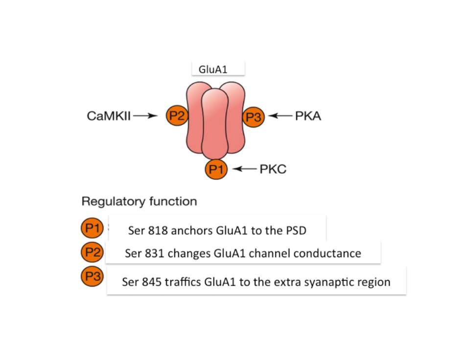
 Kinases like PKC have an inhibitory–regulatory domain and a catalytic dom...")
 As noted, PKMζ has no inhibitory domain; however, the peptide ZIP functio...")

 The activation of Trk (tyrosine kinase) receptors phosphorylates tyrosine...")


 are the inactive conformational ...")
 Schematic models of pathogenic (A) and functional (B) prions. (C) A...")


 – they remember, but there are always new filopodiu...")


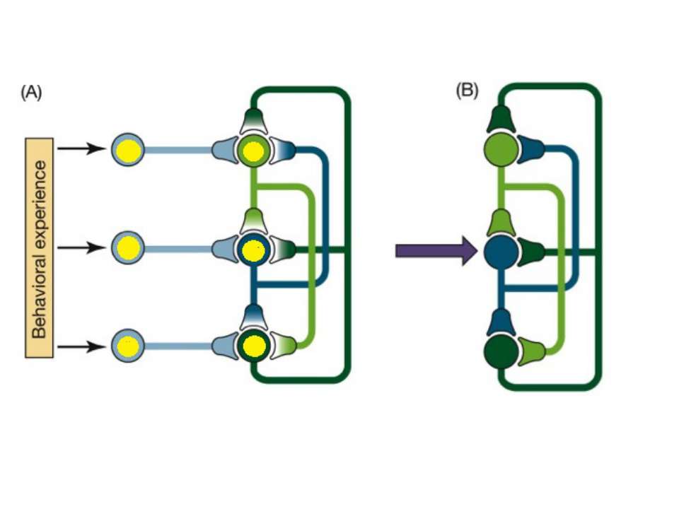
 those who fire together, wire togethe...")


")


Molecular Biology of Memory
Завантажити презентаціюПрезентація по слайдам:

In search of memory: from molecules to mind By Vasyl Mykytyuk

Acknowledgement: Professor Jerry Rudy of the psychology department at the University of Colorado at Boulder for his lectures and textbook “Neurobiology of Learning and Memory”

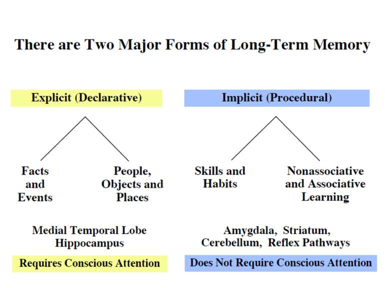
On average the human brain has 86 billion neurons Glial cells are x10 to neurons One neuron can have up to 10 000 synapses During childhood and particularly during adolescence a process “synaptic pruning” occurs – up to 50% of neurons and synapses are reduced Our brain always uses 20% of all energy

Brain functions Arousal Attention Consciousness Decision making Executive functions Natural language Learning Memory Motor coordination Perception Planning Problem solving Thought Mnemosyne (1881), mother of all Muses, goddess of memory by Dante Gabriel Rossetti Olin Levi Warner,Memory (1896). Library of Congress Thomas Jefferson Building, Washington, D.C.


The case of Henry Molaison Henry Molaison 1926-2008
")
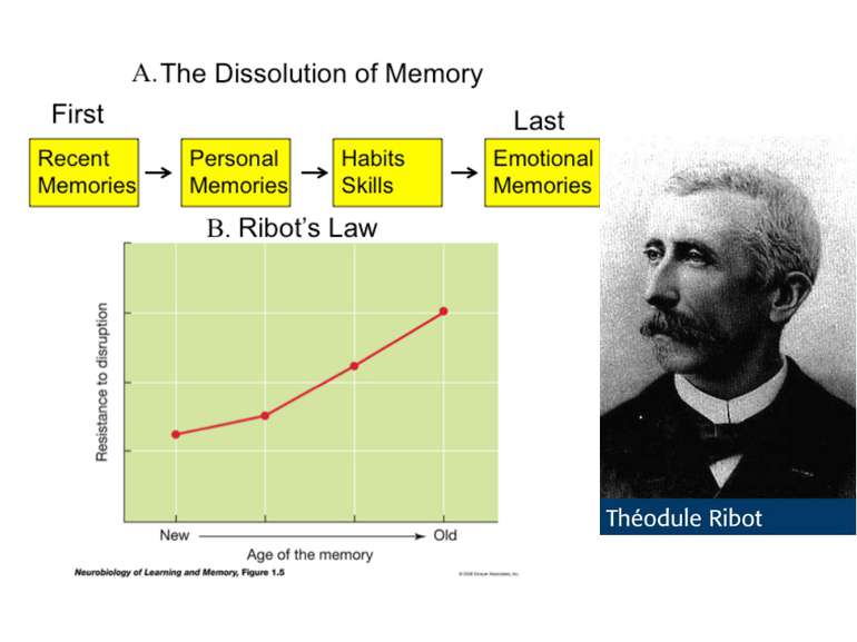
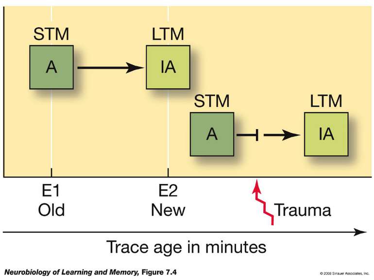
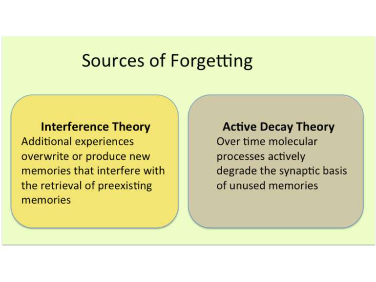
Forgetting curve by Herman Ebbinghaus (1913)

Two possible explanations of forgetting curve


Resting potential formation
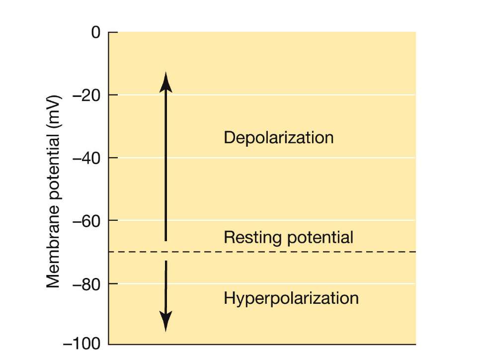

ЕЛЕКТРИЧНЕ ПРОВЕДЕННЯ ІНФОРМАЦІЇ Membrane potential Difference of charges across the plasma membrane Resting potential Resting cells are (-) inside and (+) outside Large amounts of Na+ outside the cell and K+ inside Action potential/impulse Rapid reversals in charges across the plasma membrane Caused by the exchange of ions across the membrane of the neuron Threshold level (-55mV) needed to stimulate neurons ALL-OR-NONE principle

Sequence of event during synapse: 1. Arrival of action potential to the presynaptic axon terminal 2. Neurotransmitter release from synaptic vesicles into the synaptic cleft. 3. Molecules of neurotransmitter binding to specific receptors. 4 Induction of a chemical or electrical signal in the postsynaptic cell.

The synaptic plasticity hypothesis, “the strength of synaptic connections—the ease with which an action potential in one cell excites (or inhibits) its target cell—is not fixed but is plastic and modifiable” (Squire and Kandel, 1999, p. 35).
 is produced by high-frequency stimulation (100 H...")
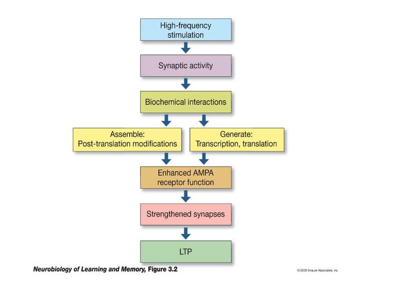
Long-Term Potentiation (LTP) is produced by high-frequency stimulation (100 Hz = 100 times in a second, three times with 20 seconds between them) Long-Term Depression (LTD) is produced by low-frequency stimulation (900 pulses of frequency 1–3 Hz ~ 15 minutes)

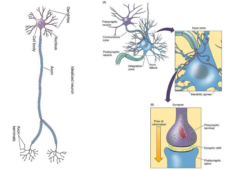
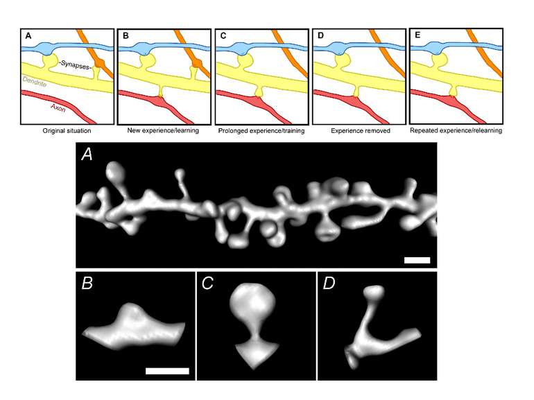
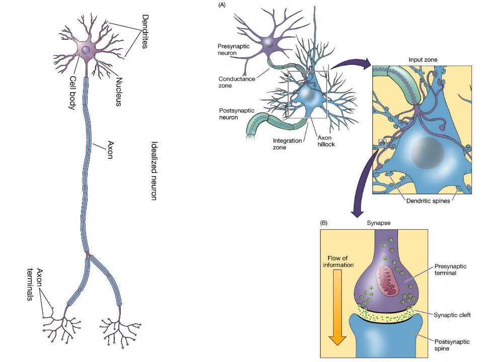
Dendrites are extensively populated with structures called dendritic spines. A spine is a small (submicrometer) membranous extrusion that protrudes from a dendrite. This figure illustrates a three dimensional reconstruction of a section of a dendrite with spines of different shapes and sizes. Dendritic spines are of special interest because (1) key receptors involved in the regulation of synaptic plasticity are located in spines and (2) changes in the composition and architecture of the spine are altered by neural activity.

 A schematic representation of an excitatory synapse. The PSD is opposed t...")
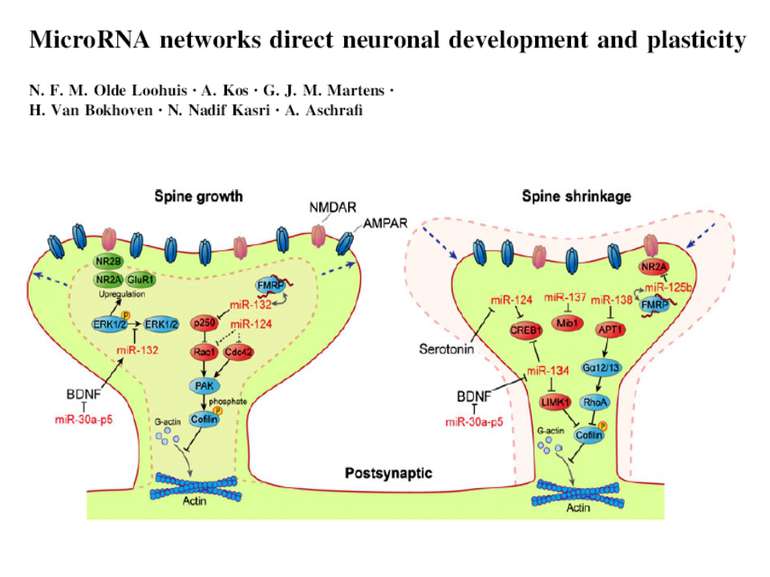
(A) A schematic representation of an excitatory synapse. The PSD is opposed to the presynaptic active zone, attached to F-actin. (B) A schematic of the PSD. Core scaffold proteins of the PSD— PSD-95, GKAP, Shank, and Homer—interact with each other and other proteins and are thought to form a lattice for the assembly of postsynaptic membrane glutamate receptors and signaling molecules.

Mechanisms of ionotropic receptor action

Two types of ionotropic glutamate receptors: The a-amino-3-hydroxyl-5-methyl-4-isoxazole-propionate (AMPA) receptor, The N-methyl-D-aspartate (NMDA) receptor They are located in dendritic spines, play a major role in the induction and expression of LTP. When gluatamate binds to these receptors their channels open and positively charged ions in the extracellular fluid (Na+ and Ca2+) enter the neuron.

APV, an NMDA receptor antagonist, prevents the induction of LTP but has no effect on its expression. CNQX, an AMPA receptor antagonist, prevents both the induction and expression of LTP.

NMDA receptor has two critical binding sites. One site binds to glutamate and the other site binds to magnesium - Mg2+. Magnesium binds to a site in the pore, forming a magnesium plug. Calcium enter requires two events: glutamate has to bind to the NMDA receptor; the synapse has to depolarize (by AMPA receptor).

The induction of LTP occurs when extracellular calcium enters the spine via the NMDA calcium channel. But entrance of calcium is not needed for LTP expression.
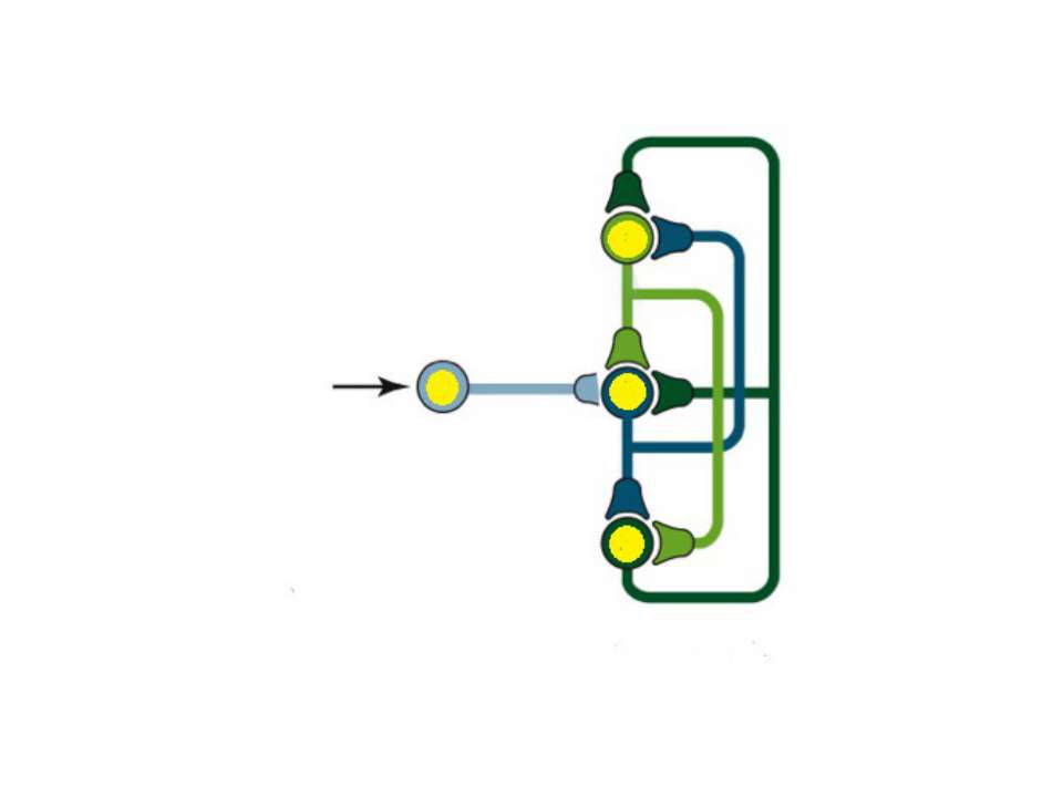
 and by synaptic ...")
AMPA trafficking is regulated constitutively (double arrows) and by synaptic activity (single arrow). The second one is a mechanism for LTP-formation.
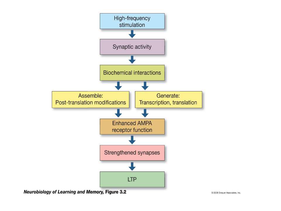

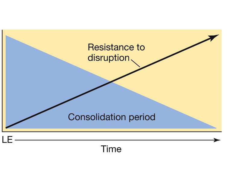
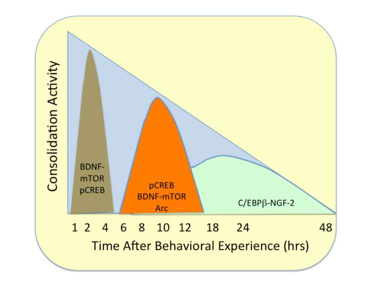
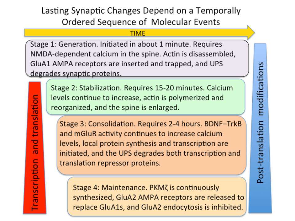
Changes in synaptic strength that support LTP evolve in stages that can be identified by the unique molecular processes that support each stage.

Generation and stabilization of trace

Constitutive trafficking of AMPA receptors
 the influx of calcium into ...")
AMPA receptors become trapped in the PSD when (1) the influx of calcium into the spine via NMDA receptors (2) activates CaMKII. This kinase phosphorylates serine (P-serine) residues on the terminal of the TARP Stargazin. This activity releases the terminal from the membrane and (3) facilitates its binding to PSD-95 complexes, thereby trapping the receptor in the PSD. The stability of the trap increases as CaMKII phosphorylates more of the Stargazin serine residues.

A.Calmodulin serves as a surrogate for second-messenger calcium and undergoes a conformational change when it binds to calcium. This new shape enables it to bind to other proteins that do not have Ca2+ binding sites. A primary target is CaMKII. B. (A) The CaMKII subunit consists of two domains, an inhibitory–regulatory domain and a catalytic domain. The regulator domain contains a phosphorylation site Thr286 that is unexposed when the kinase is in an inactive state. (B) Ca2+–calmodulin serves as a second messenger and activates the kinase. This exposes both the catalytic domain and the Thr286. Phosphorylation of this will also keep the kinase in an active state even when the second messenger dissociates from the regulatory domain. C. (A) Subunits of the kinase assemble into a ring-like complex called a holoenzyme. (B) When Ca2+–calmodulin binds to the autoinhibitory–regulatory domain of all subunits, the subunits become active and the Thr286 phosphorylation site on the regulatory unit is exposed. (C) Because of the ring-like structure of the holoenzyme, a subunit can now phosphorylate its neighbor. This process of autophosphorylation enables the subunits to remain active even when Ca2+–calmodulin is no longer present. Thus, the holoenzyme can remain in a perpetually active state.
 The prote...")
Filament actin in the spine head is crosslinked with spectrins. (B) The protease calpain is activated when calcium enters the spine through the NMDA receptors. Calpain degrades spectrins and facilitates the dissembling of actin. C. This activity contributes to the rapid insertion of additional AMPA receptors in the PSD.

Stabilizing the Trace
 (B)")
(A) (B)

When drugs such as latrunculin and cytochalasin that prevent actin polymerization are applied before TBS, they do not prevent the induction of LTP, but it rapidly decays. (B) LTP is maintained if these drugs are applied 15 minutes after LTP is induced.

Calcium entering the synapse through NMDA receptors activates two signaling cascades that regulate actin. Both pathways activate LIMK, a kinase that phosphorylates cofilin and allows actin polymerization. The second pathway, the Rac-PAK cascade, also activates processes that reorganize and crosslink actin filaments to make them resistant to depolymerization.
 Illustration of ...")
N-cadherins are reorganized by an LTP-inducing stimulus. (A) Illustration of an unpotentiated synapse with the presynaptic terminal and postsynaptic spine weakly bonded by cadherin monomers. (B) A high-frequency stimulation is delivered to this synapse to induce LTP. (C) The high-frequency stimulus promotes cadherin dimerization so the now-enlarged spine containing additional AMPA receptors is tightly coupled to the presynaptic terminal and well positioned to receive glutamate released from the presynaptic terminal.
 ...")
An LTP-inducing stimulus enhances the function of integrins in two ways. (A) Prior to induction integrins are below the plasma membrane. Integrin interacting epitopes are encrypted and the activity of metaloproteinases (MMP9s) in the extracellular matrix (ECM) is low. (B) The induction stimulus increases the expression of integrins in the spine head and the activity of MMPs in the ECM. The proteolytic activity of MMPs directed at encrypted epitopes increases availability of integrin interacting epitopes.

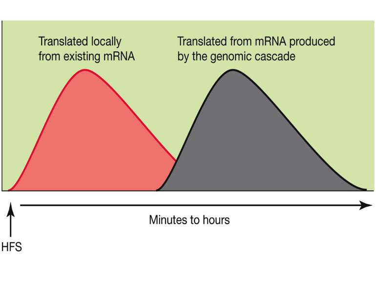
Consolidation of Synaptic Changes: Translation and Transcription

Consolidation of Synaptic Changes: Translation and Transcription New protein synthesis is essential for consolidation of synaptic changes

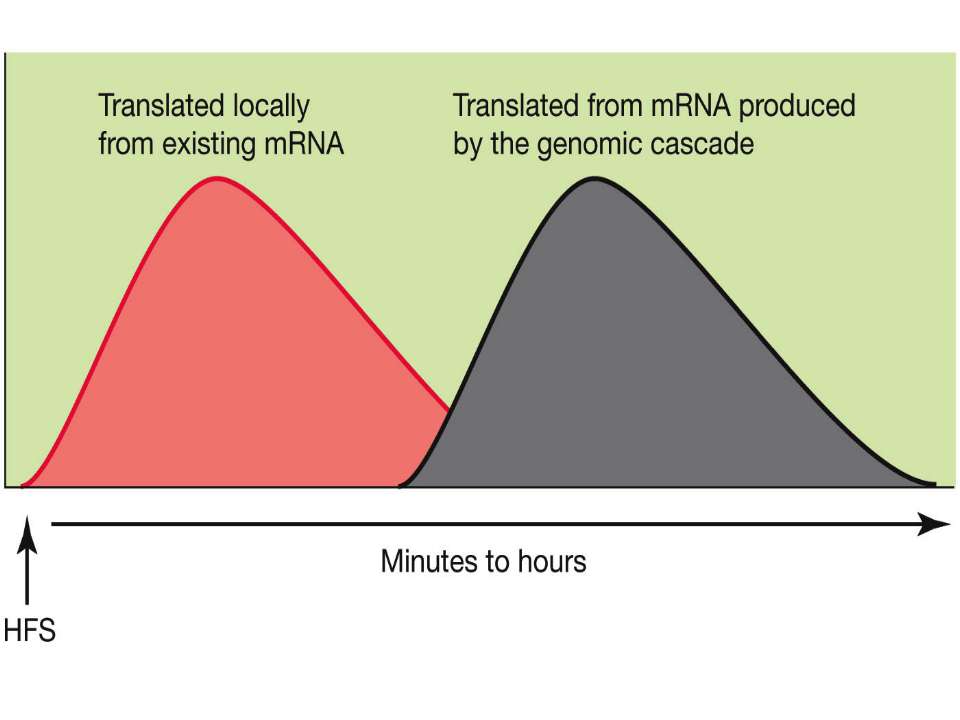
Synaptic activity simultaneously induces local mRNA translation and genomic signaling cascade, that trigger transcription of new mRNA

Messenger RNA and protein translation machinery, such as endoplasmic reticulum (ER) and ribosomes, is present locally in the dendritic spine region. Synaptic activity can activate this machinery and translate this mRNA.

Consolidation of Synaptic Changes: Specific Mechanisms The dendritic spine region called TOP(terminal oligopyrimidine tracts) has mRNAs encode for proteins such as ribosomal proteins and elongation factors that are part of the translation machinery. when synthesized these proteins enhance translation capacity in the region where they are translated. Presynaptic membrane co-releases glutamate and BDNF (brain-derived neurotrophic factor). BDNF binds to tyrosine kinase receptors, called TrkB receptors.TrkB receptors: promotes the recruitment of intracellular calcium sources 2) Activate mTOR-TOP translation 3) Arc is one of protein locally translated Regulate actin polymerization

BDNF activates local protein synthesis through PI3K/mTOR pathway
 Release of caged glutamate, coupled with a spike-time protocol, produces ...")
(A) Release of caged glutamate, coupled with a spike-time protocol, produces sustained growth and maintenance of a single spine. (B) Inhibiting the TrkB response to BDNF prevents sustained growth and maintenance. This result indicates that this protocol releases BDNF into the extracellular space.

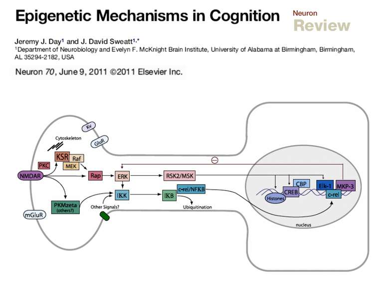
General models of how the signals reach the nucleus: synapse-to-nucleus signaling and soma-to-nucleus signaling

Synapse-to-Nucleus Signaling Different signaling cascades lead to cAMP-responsive, element-binding (CREB) protein phosphorylation and transcription initiation

Soma-to-Nucleus Signaling. Calcium enters through Voltage-Gated Calcium Channels in soma of neuron and activates calmodulin and Calcium/calmodulin-dependent protein kinase type IV which in its turn activate CREB

“cAMP Response Element-Binding Protein Is a Primary Hub of Activity-Driven Neuronal Gene Expression” Eva Benito, Luis M. Valor, Maria Jimenez-Minchan, Wolfgang Huber and Angel Barco The Journal of Neuroscience, 14 December 2011 CREB was found to occupy ≈4,000 promoter sites in vivo, but many of them are cell-specific http://in.umh-csic.es/NADtranscriptomics/tablas/CREB_regulon.txt http://in.umh-csic.es/NADtranscriptomics/tablas/CREB_timecourse.txt

Calcium entering the neuron through NMDA receptors is critical for the production of LTP but this source of calcium alone may not be sufficient to produce any form of LTP
 Calcium-induced calcium release (CICR) - extracell...")
Ryanodine receptors (RyRs) Calcium-induced calcium release (CICR) - extracellular Ca2+ enters through the NMDA receptor and binds to ryanodine receptors (RyRs) to release additional Ca2+ from the ER

Glutamate binds to a subtype of metabotropic receptor called mGluR1, which is coupled which G-protein, which in its turn activate an effector-enzyme (Phospholipase C) . When IP3 binds to IP3R calcium is released from ER in the dendritic compartment.

A single TBS train produces a short-lasting form, LTP1; four trains produce a moderately lasting form, LTP2; and eight trains produce a very long-lasting form, LTP3. • LTP1 depends only on RyRs releasing ER calcium in the dendritic spine compartment. • LTP2 depends on IP3 receptors releasing calcium from ER in the dendritic compartment. • LTP 3 depends on calcium entering the soma through vdCCs.
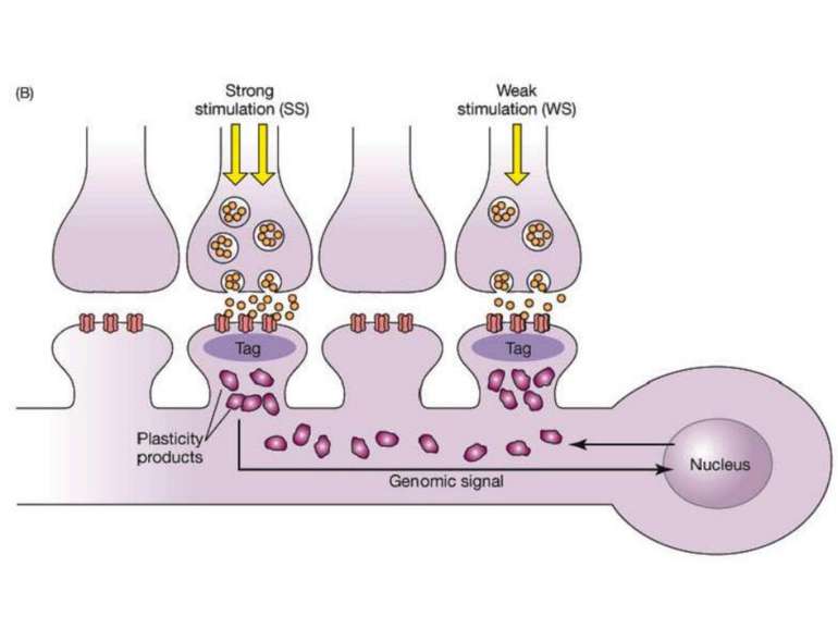
 1. Synaptic activity ...")
Synaptic tag and capture hypothesis(Frey, Morris, 1998) 1. Synaptic activity that potentiates synapses has two general effects: (a) it can generate a synaptic tag, which will allow the stimulated spine to subsequently capture newly transcribed plasticity molecules such as Arc, and (b) it can engage the translation and transcription machinery to generate new plasticity products (PPs). 2. Relatively weak stimulation (such as that needed to produce a short-lasting LTP) can create synaptic tags but will not engage the translation and transcription machinery. 3. Relatively strong stimulation will create synaptic tags and also engage the translation and transcription machinery. 4. Newly generated PPs can be captured by any tagged synapses but untagged synapses are not eligible to receive new PPs. 5. Over time, synapses lose their tag and return to their initial state. 6. Over time, the supply of new PPs will deplete. 7. To capture the PPs, the tags and new PPs must overlap in time. How do mRNAs transcribed in response to synaptic activity find their way to the correct synapses? Synaptic tag and capture hypothesis(Frey, Morris, 1998)


What is the molecular basis of the synaptic tag? Changes in functional and structural properties of spines that depend on actin filaments are a major component of the tagging operation. Potentiated spines contain more AMPA receptors and an enlarged actin cytoskeleton. Enlarged spines are better traps or tags for capturing new mRNAs/proteins needed for consolidation. One possibility is that actin-associated proteins interact with microtubules to cause them to invade the spine and permit motor proteins to cargo the relevant mRNAs/proteins to the spines that have been potentiated. In addition, once consolidated, spines with an enlarged actin cytoskeleton might successfully compete for the molecules that are needed to self-maintain and thus provide part of the solution to the molecular turnover problem.

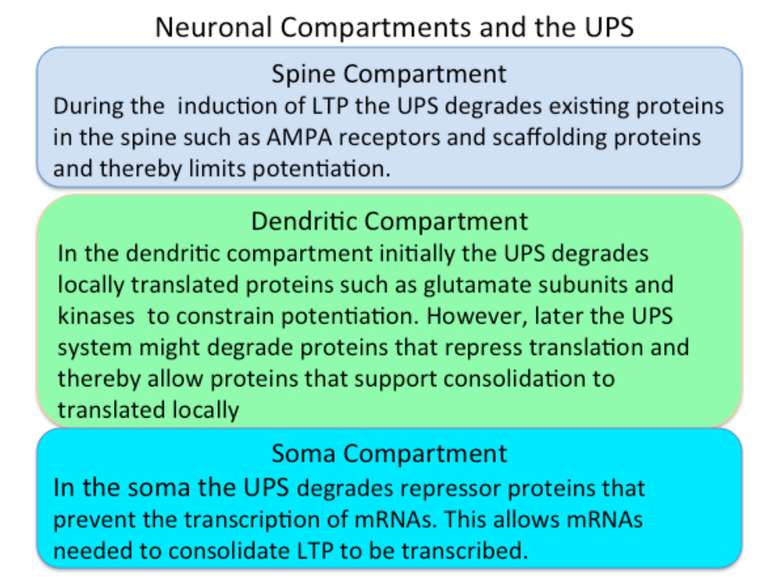
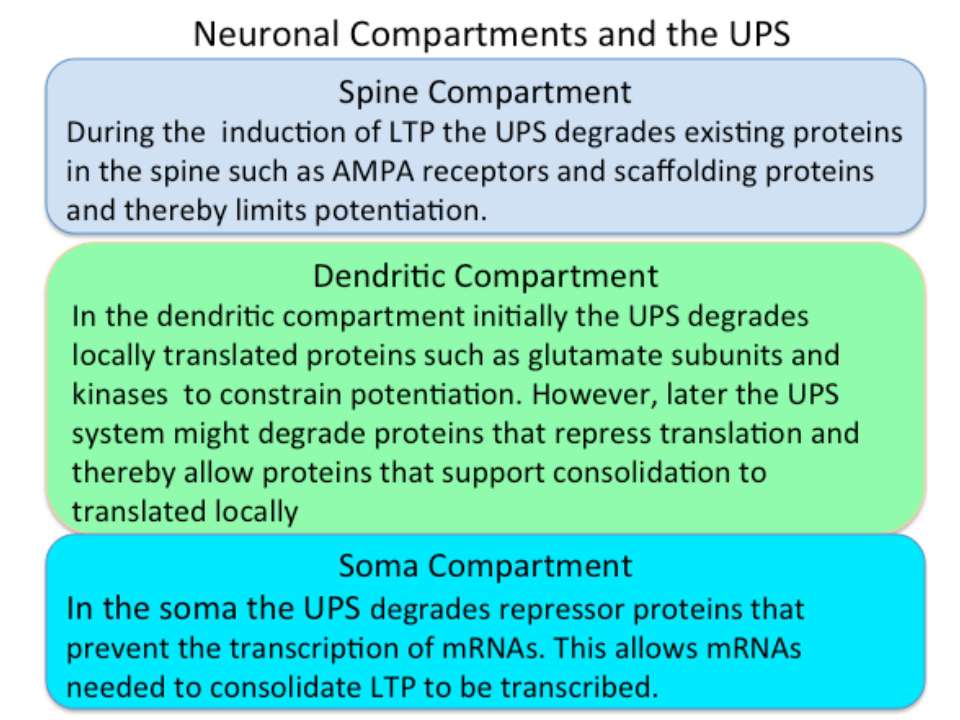
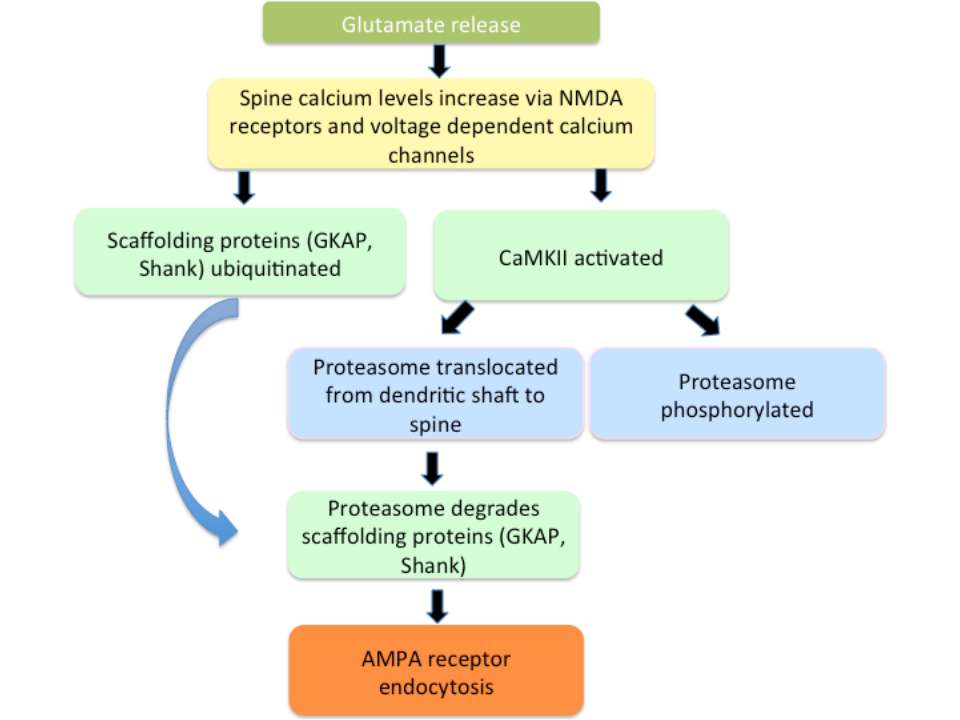
The degradation of existing proteins is important for LTP production. Components of the UPS are localized in dendrites, synapses, and the nucleus. Glutamate transmission activates the ubiquitination processes and the activation of CaMKII plays a critical role in recruiting proteasome into dendritic spines. Moreover, stimulation that induces LTP activates this system and results in protein degradation Protein Degradation Influences Three Phases of LTP: (a) an early stage that involves only post-translation modifications, (b) a later stage that depends on local protein synthesis in dendritic-spine regions, and (c) a second wave of proteins synthesized from new mRNA that is a product of genomic signaling cascades

SUMMARY OF CONSOLIDATION PROCESS

Maintaining the Consolidated Trace

Maintaining the Consolidated Trace the molecular turnover problem. The synaptic molecules that support memory traces are shortlived in comparison to the duration of our memories. Crick, however, suggested a general answer: “… the molecules in synapses interact in such a way that they can be replaced with new material, one at a time, without altering the overall state of the structure.”
 An unpotentiated synapse is depolarized. (B) As the synapse is potentiate...")
(A) An unpotentiated synapse is depolarized. (B) As the synapse is potentiated, AMPA receptors are added to the postsynaptic density. (C) Two processes—(1) degradation of AMPA receptors and (2) receptor endocytosis — then pressure the synapse to (D) return to its pre-potentiated state.
 Kinases like PKC have an inhibitory–regulatory domain and a catalytic dom...")
(A) Kinases like PKC have an inhibitory–regulatory domain and a catalytic domain. Such kinases require a second messenger (SM) to remove the inhibitory domain and expose the catalytic domain. (B) PKMζ has no inhibitory domain so it does not require second messenger to be catalytic. It is perpetually active. (C) PKMζ mRNA is present in dendritic spine regions and its translation requires synaptic activity to activate several kinases and actin filaments. (D) Once translated, PKMζ self perpetuates; it interacts with other proteins to remove the translator repressor and thereby facilitates translation of PKMζ mRNA in dendritic spines where it was initially translated.
 As noted, PKMζ has no inhibitory domain; however, the peptide ZIP functio...")
(A) As noted, PKMζ has no inhibitory domain; however, the peptide ZIP functions as an inhibitory domain and inactivates it. (B) ZIP applied to a slice prior to the inducing stimulus does not affect the early induction phase of LTP but does prevent the late phase. (C) ZIP applied 2 or 5 hours following induction reverses late-phase LTP. (After Serrano et al., 2005.)

A pool of GluA2 AMPA receptors is trapped outside of the synapse by protein interacting with C kinase 1 (PICK1). Release of this pool depends on a trafficking protein enzyme, NSF. When PKMζ interacts with the NSF–PICK1 complex, the receptors are released for entry into the PSD.
 The activation of Trk (tyrosine kinase) receptors phosphorylates tyrosine...")
(A) The activation of Trk (tyrosine kinase) receptors phosphorylates tyrosine sites on GluA2 AMPA receptors. This activity releases the receptors from the PSD into the endocytotic zone where they bond with the NSF–PICK1 complex for recycling. (B) The presence of PKMζ disrupts the normal endocytic cycle—the receptors remain trapped in the PSD and the synapse remains potentiated.

PKMζ maintains potentiated synapses through two parallel and complementary actions: (1) it facilitates the release of nonsynaptic pools of GluA2 receptors to replace GluA1 receptors and (2) it interferes with the endocytic cycle that normally removes GluA2s from the synapse.

Once synaptic activity induces PKMζ synthesis in a spine compartment (spine 2 above) in response to synaptic activity, continued synthesis could lead to PKMζ spreading from the original compartment to neighboring spines (1 and 3), where it could potentiate these spines by releasing GluA2 receptors (not shown). The spread of PKMζ is minimized because once the GluA2 receptors are released into the spine (1) they are free to recapture the PKMζ–PICK1 complex (2). In principle this autotagging process traps PKMζ in the spines where it was synthesized. Because PKMζ disrupts the endocytic cycle, an additional outcome is that GluA2s remain in the PSD region and the spine remains potentiated.

Mice were genetically modified to lack PKMζ. However, established LTP was maintained and, moreover, ZIP reversed the LTP maintenance. These unexpected results support three conclusions: (1) enduring memories and LTP can be established independent of the presence of PKMζ, that is, the maintenance of LTP does not require PKMζ, (2) ZIP is not selective for PKMζ as it also interacts with other molecules, and (3) there must be other kinases or other molecules that can participate in the maintenance of LTP and memories. There are at least two alternative explanations of these data. • PKMζ is the primary memory maintenance molecule in normal rats, and the genetic studies have uncovered redundant compensatory molecules whose contribution is masked by the normal presence of PKMζ. • PKMζ is not involved in the maintenance of LTP and maintenance depends on other kinase targets of ZIP. The field will now need to answer several questions: • What are these new targets of ZIP that must be engaged to support maintenance? • Are the processes these new targets regulate redundant with PKMζ? • What if any role does PKMζ play in maintenance? Until these questions are answered it may be appropriate to conclude that PKMζ and other kinase targets of ZIP maintain the synaptic changes that support LTP.
 are the inactive conformational ...")
The newly synthesized CPEBs (conformation A) are the inactive conformational state of the protein. Some of the protein in conformation A, either spontaneously or in a regulated way, converts into the dominant, self-perpetuating active conformation B. Few molecules in conformation B have the ability to convert all of conformation A to that of conformation B. The protein in conformation B can activate the translationally inactive mRNAs by elongating their polyA tail. The CPEB mRNA itself has a putative CPE element. Thus, once activated, the conformation B proteins can potentially regulate the availability of the proteins in conformation A. This can lead to a self-sustaining, autoregulatory feedback loop that could contribute to the stabilization of learning-related synaptic growth and the persistence of memory storage. From Bailey, C.H., Kandel, E.R., and Si, K. Neuron 2004. Prion-like protein (cytoplasmic polyadenylation element binding protein (CPEB)) is a candidate for maintenance molecule
 Schematic models of pathogenic (A) and functional (B) prions. (C) A...")
(A and B) Schematic models of pathogenic (A) and functional (B) prions. (C) Antibody that is specific for the aggregated (functional prionic) form of ApCPEB selectively blocks the maintenance of long-term facilitation produced by 5HT.

Large spines become resistant to modifications by calcium efflux out of the neck
 – they remember, but there are always new filopodiu...")
Some spines are large (A) – they remember, but there are always new filopodium-like spines (C) grow to find synapses-contact, thus there can not be problem of dendritic saturation with stable spines

Epigenetics: our experience can affect our DNA

Memory formation involves epigenetic mechanisms,

* 5-AZA – 5-azacytidine


Epigenetics: The sins of the father The roots of inheritance may extend beyond the genome, but the mechanisms remain a puzzle. Virginia Hughes

Every day we change our brain, that`s why we will never be the same tomorrow…

Molecular mechanisms of forgetting
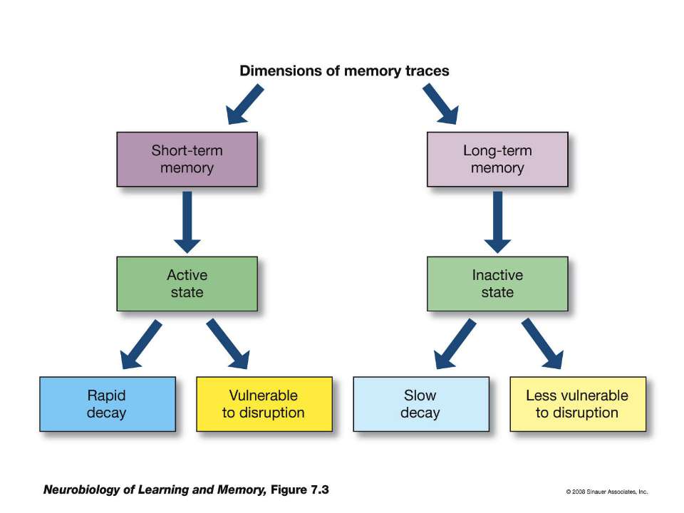

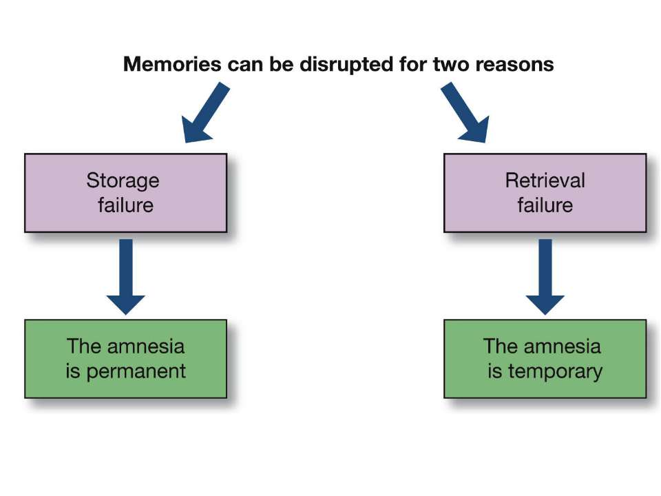
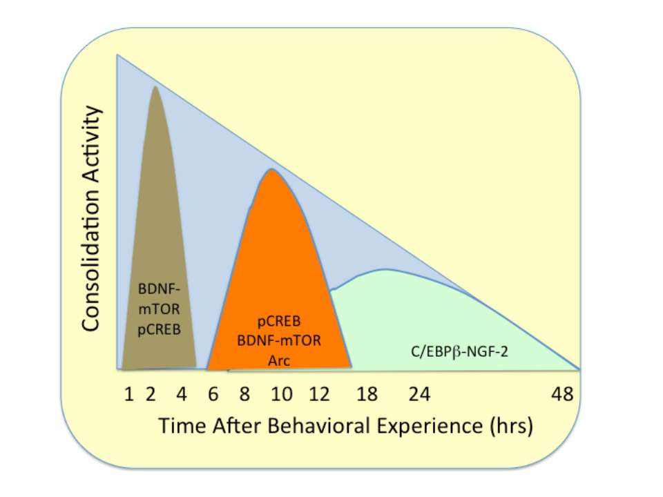

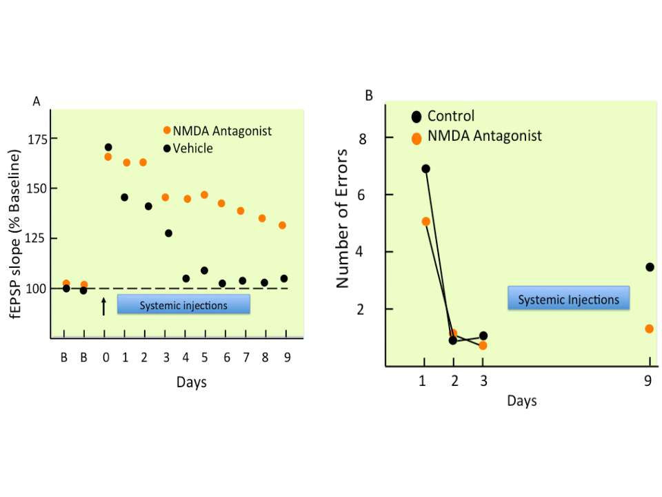

Figure 7. Model of Dopamine-Mediated Learning and Forgetting The left panel illustrates dopamine-mediated learning. Upon electric shock, a US signal is delivered when DANs release large amounts of dopamine (red spheres) onto the MBs. Binding of dopamine to the dDA1 receptor (green) leads to increased cAMP, while the CS odor stimuli results in increased Ca2+ signaling. Coincident Ca2+ and cAMP signaling leads to strong memory formation within the MBs (Tomchik and Davis, 2009). The right panel illustrates dopamine-mediated forgetting. After learning, a low level of ongoing dopamine release is sensed by the DAMB receptor (red) and weakens the memory over time. Differences in affinity of the two receptors for ligand, compartmentalization, coupling to downstream signaling pathways, or signaling kinetics may account for the dominant action of dDA1 in acquisition and DAMB in forgetting. 2012
 those who fire together, wire togethe...")
Spike-timing-dependen-plasticity (STDP) those who fire together, wire together NMDA receptor activation through depolarization after presynaptic activation results in enhancement, NMDA receptor activation through depolarization before presynaptic activation results in weakening of synaptic strength, both thought to be mediated by different levels of calcium [34].

Memory modulation

NMDA receptor can include either GluN2A or GluN2AB Receptors with GluN2AB take more time to close, so much more Calcium is influxed During our maturation our GluN2AB are replaced by GluN2A, so it is more difficult to strenghten our synapses Joe Tsien genetically engineered mice, overexpressing GluN2AB in hippocampus and cortex.

Gary Lynch and his colleagues have developed a class of drugs called ampakines that may enhance cognitive function, AMPAr are also critical for retrieval Sunifiram, Unifiram

Memory modulation framework

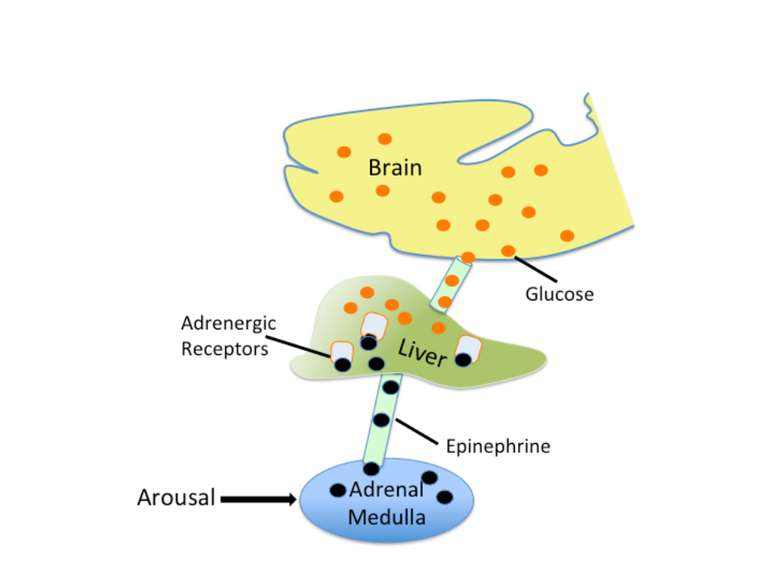
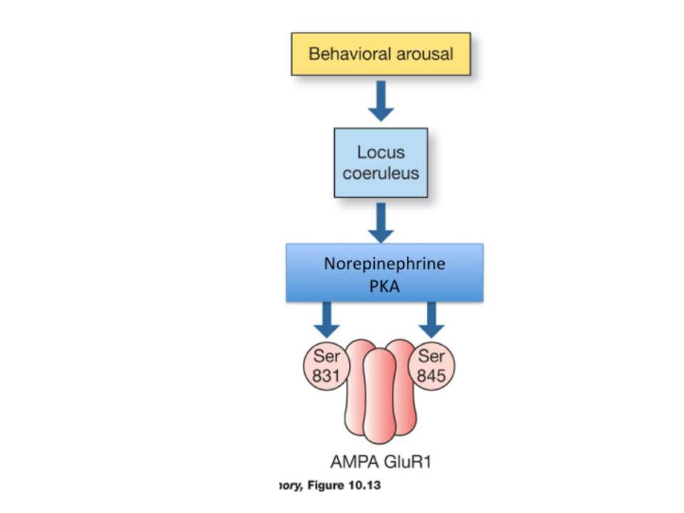
Epinephrine does not cross the blood brain barrier. However, when it is released from the adrenal medulla it binds to adrenergic receptors on the vagal nerve. In response to activation, the vagal nerve releases glutamate on neurons in the solitary tract nucleus (NTS). Activated NTS neurons release glutamate onto neurons in the locus coeruleus, which in turn release norepinephrine that binds to adrenergic receptors in the basolateral amygdala (BLA). Disrupting any component of this neuro-hormonal circuit will prevent arousal from enhancing memory.


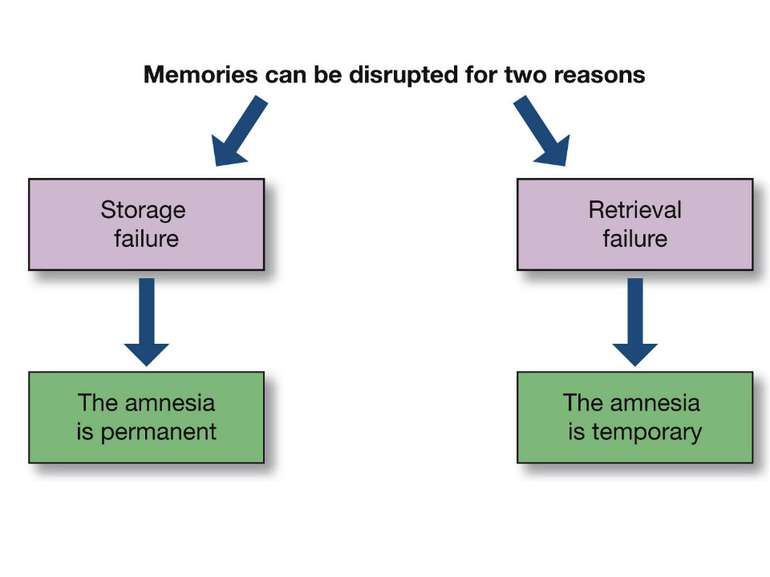
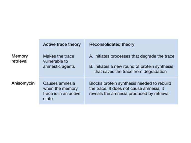
The Fate of Retrieved Memory

retrieving or reactivating the memory can return it to an active, labile state

Active trace theory

Reconsolidation theory the synapses underlying the trace become unbound or weakened retrieval also initiates another round of protein synthesis so that the trace is “reconsolidated.”

")
Model representation of reconsolidation cascade(Tronson, Nature reviews, 2007)


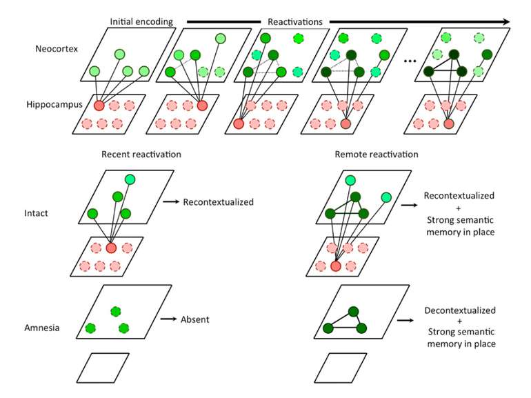
Multiple memory systems Hierarchy of Episodic memory system

The indexing theory of hippocampus role in Episodic memory

Hippocampus against entropy

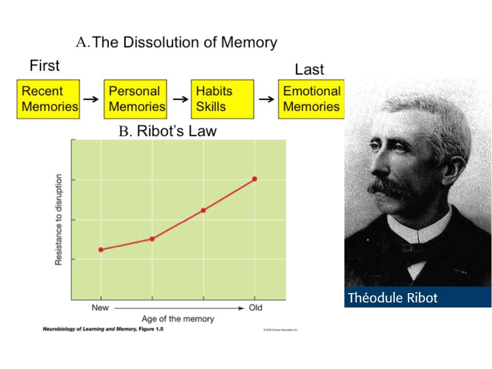
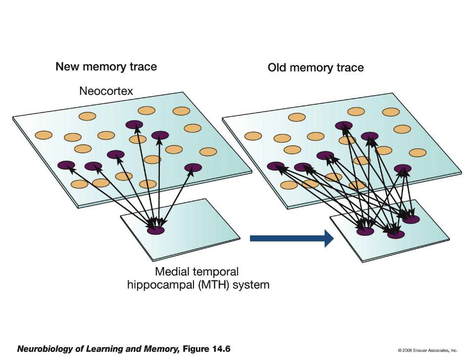
Ribot`s Law explanation


Frederick Bartlett`s serial reproduction


Memory during sleep

2004 Ribeiro, Nicolelis, 2004

Memory manipulations

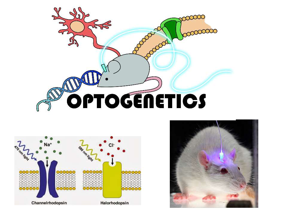
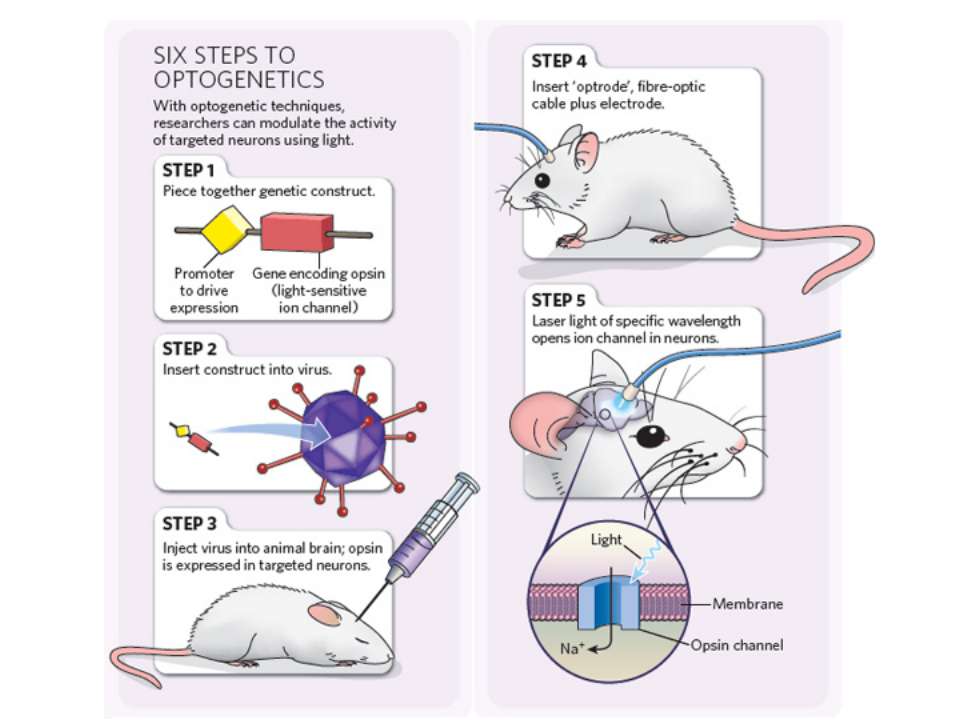

Nature, 01 June 2014

2014

Іноді достатньо згадати те, що викликає неприємні емоцї в іншому контексті. Адже згадуючи, ми кожного разу переписуємо свої спогади Пам’ять – це не те, що сталося з нами колись. Пам’ять – це те, що ми є зараз. В нашому мозку не зберігаються всі “документи” нашого життя. В ньому зберігається лише остання копія.

Схожі презентації

Категорії